SediVue Dx Urine Sediment Analyzer
The new standard for in-house urine sediment analysis
Powered by 3 cutting-edge technologies to deliver reliable results
Neural Network 4.0
- Leverages the collective learnings of 175 million patient images from IDEXX SmartService Solutions to guide confident medical decisions.
- Uses advanced Neural Network 4.0 algorithmic software and machine-learning capabilities that enable the analyzer to better identify abnormalities with each result generated.
Inverted microscope with built-in camera
- Examines the equivalent of 45 high-power fields from beneath the sample to maximize visibility of clinically relevant particles.
- Captures 70 high-resolution images per run.
- Autofocuses on each shot for crisp images.
- Adjusts exposure with each image to ensure optimal contrast.
Purpose-built onboard gravitational centrifuge
- Spins at a significantly lower force than most in-house centrifuges to preserve delicate casts that external centrifuges break apart and to ensure sample integrity.
- In 30 seconds, gently deposits formed elements into a monolayer at the bottom of the cartridge.
High-resolution digital images
Images provide diagnostic proof in real-time
The following cases show how veterinarians used results to determine next steps quickly and confidently.
Images provide diagnostic proof in real-time
The following cases show how veterinarians used results to determine next steps quickly and confidently.
Elements Identified:
Red blood cells, white blood
cells, and bacteria
Clinical Decisions:
The veterinarian confirmed a urinary tract infection was present during the visit. A urine sample was submitted for quantitative urine culture and antimicrobial sensitivities.
Red blood cells, white blood
cells, and bacteria
Clinical Decisions:
The veterinarian confirmed a urinary tract infection was present during the visit. A urine sample was submitted for quantitative urine culture and antimicrobial sensitivities.
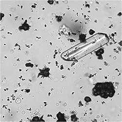
Elements Identified:
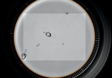
Ammonium biurate crystals
Clinical Decisions:
Liver disease was suspected, and the veterinarian followed up with a bile acids test. Abdominal radiographs revealed a small liver, and an ultrasound with color-flow Doppler confirmed the presence of a portosystemic shunt.
Ammonium biurate crystals
Clinical Decisions:
Liver disease was suspected, and the veterinarian followed up with a bile acids test. Abdominal radiographs revealed a small liver, and an ultrasound with color-flow Doppler confirmed the presence of a portosystemic shunt.
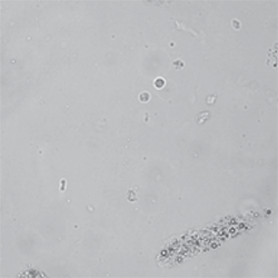
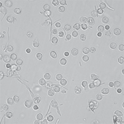
Elements Identified:
Numerous nonhyaline casts
Clinical Decisions:
Renal tubular injury was indicated, and the veterinarian recommended the IDEXX SDMA Test and a repeat urinalysis in 2 weeks.
Numerous nonhyaline casts
Clinical Decisions:
Renal tubular injury was indicated, and the veterinarian recommended the IDEXX SDMA Test and a repeat urinalysis in 2 weeks.
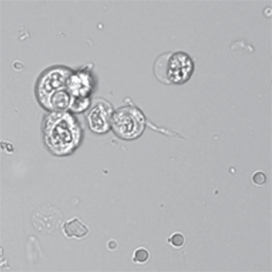
Elements Identified:
Numerous nonsquamous epithelial cells
Clinical Decisions:
The veterinarian followed up with a stained, cytological slide preparation, further classifying the cells as transitional cell carcinoma.
Numerous nonsquamous epithelial cells
Clinical Decisions:
The veterinarian followed up with a stained, cytological slide preparation, further classifying the cells as transitional cell carcinoma.
Information from Idexx.com/en/veterinary/analyzers